Navigation
Public engagement

Virus Fighter

Build a virus or fight a pandemic!

Play online

Maya's Marvellous Medicine

Read online for free

Print your own copy

Battle Robots of the Blood

Read online for free

Print your own copy

Just for Kids! All about Coronavirus

Read online for free

Print your own copy

Archive
LabListon on Twitter

Entries in immunology (100)

Tuesday
Jun142022

Greater understanding of immune signalling molecule raises hope for improved clinical use

Source of immune signals alters the immune response

Key points:

  • Researchers have identified source-specific effects of the signalling molecule interleukin 2 (IL2) on the immune response.
  • IL2 is an important signalling molecule that has been harnessed as a biologic therapy for a number of diseases but can result in unwanted side-effects.
  • This study, conducted using new mouse models, found that the immune response to IL2 is dependent on the cellular source of the IL2 production.
  • Their new insight explains the link between IL2 treatments and side-effects, opening up the potential to apply this powerful immune modulator to optimise treatments while avoiding off-target effects.

A detailed update to our understanding of the key immune system signalling molecule interleukin 2 has been published today by researchers at the Babraham Institute. Their findings explain common side effects of IL2-based therapies, and identify potential new uses of IL2 as an immune-modulating biologic drug. This research was only possible thanks to a new mouse model which allowed researchers to control which immune cell types produced IL2. With further research, this understanding of the rules dictating which cells respond to IL2 could allow scientists to optimise autoimmune and cancer treatment while avoiding unwanted side-effects.

Dr Carly Whyte, lead author on the paper who undertook this research as a postdoctoral researcher in the Liston lab, said: "IL2 is a protein that is normally tightly regulated in the immune system because it has such strong effects. However, when IL2 is given as a therapeutic treatment, these normal restrictions on IL2 are overruled. By using mouse models, we have found that the presence of IL2 in certain zones of the immune system leads to some of the same side-effects that we see in human patients treated with IL2. We hope that by understanding more about how IL2 works in different zones, this treatment might be tailored to be more effective."

IL2 is involved in a large number of different communication networks in the immune system, being produced by a variety of cellular sources and affecting a diversity of cell ‘responders’. It is not only needed for maintaining regulatory T cells, which prevent our body’s immune system from attacking itself, but also CD8 T cells, which attack tumour cells and virus-infected cells. Owing to this dual functionality, IL2 has been harnessed to both promote an immune response, and limit one, depending on the target cells. Despite being actively explored in hundreds of ongoing clinical trials, the full therapeutic potential is currently limited by frequently-encountered side-effects.

Previous explanations for these side-effects were based on the high doses of IL2 when given as a biologic drug, but Prof. Adrian Liston and his team were able to demonstrate that the cell-type making IL2, and the location of those cells, dramatically change the consequences of IL2 exposure. Dr Kailash Singh, co-lead author, explains "Our genetically modified mouse models showed that the immune responses are varied depending on the source of IL2. Our findings revealed that the IL2 response is very much context-dependent, and is not solely due to the concentration of IL2."


Prof. Liston, a senior group leader in the Institute’s Immunology research programme, said: “This work changes the way we think about IL2 as a decades-old therapeutic molecule, demonstrating that it is not just the dose of the IL2 that matters, but also where it is located in the body. Putting together the pieces of this cause and effect intricacy has involved several remarkable scientists and over a decade of research. It was only by bringing together experts in animal research, flow cytometry and immunology, that we had the know-how to tackle the complexity of this question. We’re increasingly aware of the therapeutic power of the immune system, and these findings provide a new avenue of investigation for designing biologic drugs.”

Dr James Dooley, joint senior author of the study, said: "The next generation of biologics will be smarter and tailored to the biology of the disease. This work teaches us that one route of smart design of IL2 is to target delivery to different parts of the body, potentially allowing us to drive very different therapeutic outcomes in patients."

Read the original paper at The Journal of Experimental Medicine!

Friday
Jun102022

CYTO 2022 presentation by Dr Oliver Burton

If you missed Oliver's presentation on AutoSpill at CYTO, here is his presentation on AutoSpill, in conjunction with FlowJo.


If you are interested, you can read the original paper here, or use the AutoSpill website to compensate your data here.
Wednesday
Jun012022

Drugs, Vaccines and a Hopeful Future: Exploring Advances in Multiple Sclerosis Research

I was interested by Ruairi Mackenzie from Technology Networks for World Multiple Sclerosis Day. Here is the resulting article:


World Multiple Sclerosis (MS) Day recognizes the millions of people worldwide who are affected by this neuroimmunological disease. The campaign site for World MS Day 2022 strikes an optimistic chord, seeing the date as a chance to “celebrate global solidarity and hope for the future”. This year, there is more reason to buy into that optimism that ever before.

 Recognizing the immune basis of MS

Adrian Liston, a group leader at the Babraham Institute, based near Cambridge, UK, is well-placed to explain that sunny outlook. He first studied MS as part of an undergraduate project.  Over the two decades since, Liston has continued to research in the MS field, watching science’s understanding of the disease deepen.

“I think the most profound change has been the recognition that MS is an immune-mediated disease,” says Liston. The symptoms of MS are neurological – patients experience a range of sensory and motor conditions, including fatigue, numbness, spasms and weakness and loss of control over muscle movement or function. While MS has long been thought to include an immune component – Jean Martin Charcot, the French doctor who first described the disease, noted the presence of immune cells in patients’ spinal cords – it was only  recently that the immune-mediation of the disease was fully understood. It’s now recognized that the neurological symptoms are a consequence of immune cells infiltrating the brain and attacking the myelin sheath coating that permits normal nerve cell conduction.

Part of that understanding came from the galloping pace of genetics research. The neurological symptoms of MS are in contrast with its genetic signature, explains Liston. From a geneticist’s point of view, “MS looks a lot like Type 1 diabetes, or any of these other autoimmune diseases, and the same genes are controlling it,” he says.

These genetic insights were added to a rapidly growing body of preclinical research. Experimental autoimmune encephalomyelitis (EAE) is an inflammatory autoimmune disease seen in animals that mimics the disease course of MS. This model proved essential for the development of drugs for MS. Liston explains that these drugs came in two waves. The first was heralded by the approval of interferon beta-1b in 1993, the first drug capable of altering the course of MS. The interferons, which reduce the number of immune cells crossing the blood-brain barrier, showed success at improving MS symptoms. This provided direct evidence that adopting an immune-targeting approach could help patients.

A new generation of drugs

For some, these early drugs have proved enduringly beneficial and remain their main course of treatment years later. MS drug development, however, continued to refine the targeting of the immune system. “What we really we have now is the second wave of drug generation, where we have much more sophisticated immune-modulating molecules, and we can really target very specific pathways of cells that are causing the damage,” says Liston. These new drugs (there are 23 FDA-approved mediations for MS as of 2022) work better and for longer.

Liston says that an MS patient today can expect fewer symptoms and far longer periods of good health than in the past. 85% of MS patients have a relapsing-remitting form of the disease, which sees symptoms wax and wane. While 20 years ago MS may have induced multiple relapses each year, with potentially permanent loss of function a risk each time, today a patient that responds well to the latest treatments can expect to go five or even ten years without any further relapses, says Liston: “It gives them a life well beyond diagnosis, which wasn't the case 20 years ago.”

The focus of these treatments is to minimize the damage of any immune attack on the brain. Can we also restore function lost during these attacks? Research in this area is progressing, if at a much slower rate. Data presented in 2020 showed that a compound, bexarotene (full disclosure: I’ve published research on this drug myself) was shown to restore some of the myelin lost during the disease course. Side effects of the drug and limited clinical impact meant bexarotene was not taken forward to approval. The challenges of restoring the damaged brain are significant, but this research shows that, in principle, healing the brain’s myelin might one day be possible.

A vaccine to prevent MS?

In the meantime, other findings have pointed a way to intercept MS earlier, stopping the disease in its tracks before it can ever cause damage to the brain.

It all begins with a virus.

Epstein-Barr virus (EBV), a type of herpesvirus, has long been associated with MS – studies had noted a higher risk of contracting MS after previously having infectious mononucleosis (IM), a disease caused by EBV – but a “smoking gun” had been hard to identify.

This is because up to 95% of the adult population have EBV, which is an incredibly successful virus that, after infecting a host, can lay dormant for years. Designing the right sort of study to test whether getting EBV would later increase your MS risk was therefore a huge logistical challenge. In January, however, researchers at Harvard Medical School met that challenge. Using a longitudinal method, the team collected blood serum samples from US military personnel, who are required to submit a blood serum sample at the start of and then after every two years of their service.

With samples from 10 million different people stored, the study was easily able to identify individuals who didn’t have EBV during their first sample, as well as those who developed MS during the course of their service. The study showed that, of 35 individuals who tested negative for EBV on enrolling, all but one of them went on to become infected with EBV before developing MS.

This corresponds to a 32-fold increased risk of developing MS. To put this in perspective, the strongest genetic risk factor for MS – which involves having a set of particular immune genes – confers a three-fold risk. This association is so strong, that the study, in Liston’s opinion, “finished the argument” on whether EBV plays a causal role in MS. The underlying theory is that, in some individuals, the body responds to the presence of the virus by mistakenly attacking the brain’s myelin sheath, triggering MS’s symptoms.

“Incredibly profound” implications

The finding and its implications, says Liston, are “incredibly profound”: “I'd say the best analogy is of human papilloma virus (HPV) and cervical cancer. Cervical cancer and some anal cancers as well are largely caused by infections with the virus, HPV. Now that knowledge was very controversial for a long time.”

The evidence linking HPV and cervical cancer is now solid, Liston says: “Most people who have the virus never get the cancer. But what that allows us to do is then go and develop vaccines for HPV. By preventing the infection, you essentially prevent the cancer formation.”

If a similar approach could be taken with EBV – vaccinating every child against the virus before they are infected – future generations could essentially be protected from ever acquiring MS. That’s a hugely exciting prospect. There is much more research to be done, however, before that reality is met. Liston points out that EBV “is not a trivial virus to attack” – it tends to hide within patients’ B cells and is happy to remain concealed there for a patient’s lifetime. Much more work will need to be conducted on how to target EBV and on the steps between infection and symptoms of MS.

The future of MS research

For now, says Liston, anyone interested in following the progress of the MS field should pay special attention to studies that improve the personalization of existing treatments. “We really want to be able to match up which medication is going to work on which person. In the case of MS, that is probably the single most important thing,” he explains. Currently, patients can endure multiple relapses while testing out which treatment works best for them. Work into matching patients with certain backgrounds and disease progressions with an appropriate drug could be hugely significant, says Liston.

The other exciting advance he mentions is the creation of better targeted drugs. Despite the advances of second-generation MS drugs, Liston says that in terms of specificity, they are essentially immune sledgehammers.

“In someone with MS, only 0.1% of their white blood cells are dangerous,” he says. But current treatments hit that 0.1% alongside a vast swathe of the rest of the immune system, which can have extreme side effects. New generations of drugs, Liston explains, will be better targeted at immune culprits. One field, antigen-specific tolerance work, looks to suppress the incorrect response by confused immune cells, restoring the balance of the immune system without impairing it from its important role of protecting our body against outside threats.

Time to be excited about the present

Is Liston hopeful about the future of MS research? “It’s not even just the future,” he responds, “I’m actually quite positive about the present of MS compared to where we were 20 years ago.”

The promise that drugs targeting the immune system could help MS patients has been well met. “We're not talking about cold fusion, where they've been promising boundless energy in 10 years’ time, and promising that for 40 years, we’re talking about a place where the promises have been delivered over and over,” says Liston. He believes that patients are going to continue to have longer, healthier lives as drugs improve and become smarter and more targeted.

The only note of caution Liston sounds is in managing our expectations: “We're not going to have these type of ‘Eureka!’ moments where there's one tablet that just cures MS, but we will be coming closer and closer to a place where there will be a treatment regime that works on almost every patient and is almost perfect.”

Those advances will partly come from innovative use of the latest techniques – Liston’s lab recently published work where gene therapy was used to edit a small fraction of cells in the brain. The edit caused these cells to produce a pro-survival molecule in the surrounding brain area. This is turn helped increase the numbers of regulatory T cell (Tregs) – a cell able to suppress damaging immune responses – in the brain. In animal models, the longer-living Tregs were able to not only improve MS-like symptoms but facilitate quicker healing from brain injury.

Neurological disease can so often seem intractable, beset by the complications of the brain and our still juvenile understanding of how the biological wonders in our heads function and fail. But on World MS Day 2022, there is plenty good news, at least in one small corner of brain medicine. “This is a very hopeful time for patients,” Liston concludes.

Thursday
May262022

Harnessing the immune system to treat traumatic brain injury 

Pioneering new treatment leads to improved recovery from brain trauma in mice


Research offers new approach to limiting harmful brain inflammation after injury or disease

  • Researchers have designed a targeted therapeutic treatment that restricts brain inflammation. The effectiveness of this approach in improving outcomes was demonstrated following brain injury, stroke or multiple sclerosis in mice.
  • The system increases the number of regulatory T cells, mediators of the immune system’s anti-inflammatory response, in the brain.
  • By boosting the number of T regulatory cells in the brain, the researchers were able to prevent the death of brain tissue in mice following injury and the mice performed better in cognitive tests.
  • The treatment has a high potential for use in patients with traumatic brain injury, with few alternatives currently available to prevent harmful neuroinflammation.

A therapeutic method for harnessing the body’s immune system to protect against brain damage is published today by researchers from the Babraham Institute’s Immunology research programme. The collaboration between Prof. Adrian Liston (Babraham Institute) and Prof. Matthew Holt (VIB and KU Leuven; i3S-University of Porto) has produced a targeted delivery system for boosting the numbers of specialised anti-inflammatory immune cells specifically within the brain to restrict brain inflammation and damage. Their brain-specific delivery system protected against brain cell death following brain injury, stroke and in a model of multiple sclerosis. The research is published today in the journal Nature Immunology.

Traumatic brain injury, like that caused during a car accident or a fall, is a significant cause of death worldwide and can cause long-lasting cognitive impairment and dementia in people who survive.  A leading cause of this cognitive impairment is the inflammatory response to the injury, with swelling of the brain causing permanent damage. While inflammation in other parts of the body can be addressed therapeutically, but in the brain it problematic due to the presence of the blood-brain barrier, which prevents common anti-inflammatory molecules from getting to the site of trauma.

Prof. Liston, a senior group leader in the Institute’s Immunology programme, explained their approach: “Our bodies have their own anti-inflammatory response, regulatory T cells, which have the ability to sense inflammation and produce a cocktail of natural anti-inflammatories. Unfortunately there are very few of these regulatory T cells in the brain, so they are overwhelmed by the inflammation following an injury. We sought to design a new therapeutic to boost the population of regulatory T cells in the brain, so that they could manage inflammation and reduce the damage caused by traumatic injury.”

The research team found that regulatory T cell numbers were low in the brain because of a limited supply of the crucial survival molecule interleukin 2, also known as IL2. Levels of IL2 are low in the brain compared to the rest of the body as it can’t pass the blood-brain barrier.

Together the team devised a new therapeutic approach that allows more IL2 to be made by brain cells, thereby creating the conditions needed by regulatory T cells to survive. A ‘gene delivery’ system based on an engineered adeno-associated viral vector (AAV) was used: this system can actually cross an intact blood brain barrier and deliver the DNA needed for the brain to produce more IL2 production.

“Even though the number of Treg cells in the naive CNS is very low, they are a powerful immunosuppressive tool that we exploited  to reduce damage-induced inflammation without harnessing the brain.” explains the first author, Dr. Pasciuto.

Commenting on the work, Prof. Holt said ‘For years, the blood-brain barrier has seemed like an insurmountable hurdle to the efficient delivery of biologics to the brain. Our work, using the latest in viral vector technology, proves that this is no longer the case; in fact, it is possible that under certain circumstances, the blood-brain barrier may actually prove to be therapeutically beneficial, serving to prevent ‘leak’ of therapeutics into the rest of the body’.

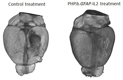
The new therapeutic designed by the research teams was able to boost the levels of the survival molecule IL2 in the brain, up to the same levels found in the blood. This allowed the number of regulatory T cells to build up in the brain, up to 10-fold higher than normal. To test the efficacy of the treatment in a mouse model that closely resembles traumatic brain injury accidents, mice were given carefully controlled brain impacts and then treated with the IL-2 gene delivery system. The scientists found that the treatment was effective at reducing the amount of brain damage following the injury, assessed by comparing both the loss of brain tissue and the ability of the mice to perform in cognitive tests.

Lead author, Dr Lidia Yshii, explained: “Seeing the brains of the mice after the first experiment was a ‘eureka moment’ – we could immediately see that the treatment reduced the size of the injury lesion”.

Recognising the wider potential of a drug capable of controlling brain inflammation, the researchers also tested the effectiveness of the approach in experimental mouse models of multiple sclerosis and stroke. In the model of multiple sclerosis, treating mice during the early symptoms prevented severe paralysis and allowed the mice to recover faster. In a model of stroke, mice treated with the IL2 gene delivery system after a primary stroke were partially protected from secondary strokes occurring two weeks later.  In a follow-up study, still undergoing peer review, the research team also demonstrated that the treatment was effective at preventing cognitive decline in ageing mice.

“By understanding and manipulating the immune response in the brain, we were able to develop a gene delivery system for IL-2 as a potential treatment for neuroinflammation. With tens of millions of people affected every year, and few treatment options, this has real potential to help people in need. We hope that this system will soon enter clinical trials, essential to test whether the treatment also works in patients." said Prof. Liston.

Dr Ed Needham, a neurocritical care Consultant at Addenbrooke’s Hospital who was not a part of the study, commented on the clinical relevance of these results: “There is an urgent clinical need to develop treatments which can prevent secondary injury that occurs after a traumatic brain injury. Importantly these treatments have to be safe for use in critically unwell patients who are at high risk of life-threatening infections. Current anti-inflammatory drugs act on the whole immune system, and may therefore increase patients' susceptibility to such infections. The exciting progress in this study is that, not only can the treatment successfully reduce the brain damage caused by inflammation, but it can do so without affecting the rest of the body's immune system, thereby preserving the natural defences needed to survive critical illness."

Thursday
May262022

Baanbrekende nieuwe behandeling leidt tot beter herstel na hersentrauma bij muizen

Onderzoekers hebben een gerichte therapie ontworpen die ontsteking in de hersenen tegengaat. De aanpak – waarbij doelgericht DNA tot in de hersenen wordt gebracht - blijkt succesvol bij muizen met een hersenletsel, beroerte of multiple sclerose

Kort & bondig:

  • Onderzoekers hebben een gerichte therapie ontworpen die ontsteking in de hersenen tegengaat. De aanpak – waarbij doelgericht DNA tot in de hersenen wordt gebracht - blijkt succesvol bij muizen met een hersenletsel, beroerte of multiple sclerose. 
  • De behandeling bestaat er in het aantal regulatoire T-cellen, die de anti-inflammatoire respons van het immuunsysteem reguleren, te verhogen in de hersenen.  
  • Door het aantal regulerende T-cellen in de hersenen te verhogen, konden de onderzoekers het afsterven van hersenweefsel bij muizen na verwonding voorkomen en deden de muizen het beter bij cognitieve testen. 
  • Voor patiënten met een traumatisch hersenletsel zijn er momenteel weinig opties om schadelijke neuroinflammatie te voorkomen. De nieuwe resultaten zijn dus erg hoopgevend. 

Onderzoekers uit Leuven en uit het Verenigd Koninkrijk publiceren vandaag nieuwe resultaten over een therapeutische piste waarbij het immuunsysteem wordt ingezet om hersenbeschadiging te voorkomen. De samenwerking tussen professor Adrian Liston (voorheen VIB en KU Leuven en sinds enkele jaren verbonden aan het Babraham Institute in het VK) en professor Matthew Holt (verbonden aan VIB, KU Leuven en de i3S-Universiteit van Porto) leidde tot een systeem om in de hersenen het aantal gespecialiseerde ontstekingsremmende immuuncellen op te voeren om op die manier hersenontsteking en -beschadiging te beperken. De aanpak bleek alvast in muismodellen tot minder hersenschade te leiden na hersenletsel, beroerte of bij multiple sclerose. Het onderzoek wordt vandaag gepubliceerd in het tijdschrift Nature Immunology. 

Traumatisch hersenletsel, zoals veroorzaakt na een auto-ongeval of een val, is wereldwijd een belangrijke doodsoorzaak. Het kan langdurige en ernstige gevolgen hebben voor mensen die het overleven, onder de vorm van cognitieve problemen en zelfs dementie. Een belangrijke oorzaak van deze cognitieve stoornissen is de ontstekingsreactie op het letsel. Hierbij veroorzaakt zwelling van de hersenen permanente schade. Terwijl ontsteking in andere delen van het lichaam kan worden aangepakt met geneesmiddelen, is dit in de hersenen heel moeilijk door de aanwezigheid van de bloed-hersenbarrière, die voorkomt dat gewone ontstekingsremmende moleculen op de plaats van het (hersen)trauma kunnen komen.  

Prof. Liston: "Ons lichaam heeft zijn eigen ontstekingsremmende respons: regulatoire T-cellen zijn in staat om ontstekingen waar te nemen en een cocktail van natuurlijke ontstekingsremmers te produceren. Helaas zijn er maar heel weinig van deze regulerende T-cellen in de hersenen. Wij probeerden een nieuw therapeutisch middel te ontwikkelen om de hoeveelheid regulerende T-cellen in de hersenen te vergroten. Indien er voldoende regulatoire T-cellen zijn, zo redeneerden we, zouden ze de ontsteking na een letsel kunnen beheersen en de schade beperken." 

Het onderzoeksteam ontdekte dat het aantal regulerende T-cellen in de hersenen laag was door een beperkte aanvoer van het cruciale overlevingsmolecuul interleukine 2, ook bekend als IL2. Het niveau van IL2 is laag in de hersenen vergeleken met de rest van het lichaam omdat het niet doorheen de bloed-hersenbarrière kan.  

Samen bedacht het team een nieuwe therapeutische aanpak waardoor meer IL2 kan worden aangemaakt door hersencellen. De onderzoekers gebruikten een "gene delivery"-systeem op basis van een virale vector: dit systeem kan daadwerkelijk een intacte bloed-hersenbarrière passeren en het DNA afleveren dat de hersenen nodig hebben om meer IL2 aan te maken.  

Prof. Holt: "Jarenlang leek de bloed-hersenbarrière een onoverkomelijke hindernis voor de efficiënte toediening van biologische geneesmiddelen in de hersenen. Ons werk, waarbij we gebruik maken van de nieuwste virale vectortechnologie, bewijst dat dit niet langer het geval is; het is zelfs mogelijk dat de bloed-hersenbarrière onder bepaalde omstandigheden therapeutisch gunstig kan zijn, omdat ze – eenmaal op hun bestemming – het 'lekken' van geneesmiddelen naar de rest van het lichaam verhindert." 

Dankzij hun aanpak waren de onderzoekers in staat de niveaus van de overlevingsmolecule IL2 in de hersenen op te voeren tot dezelfde niveaus als in het bloed. Hierdoor kon het aantal regulerende T-cellen zich in de hersenen opbouwen, tot 10 maal hoger dan normaal. Om de doeltreffendheid van de behandeling te testen in muizen. Wat bleek? Muizen met meer IL2 hadden inderdaad minder hersenschade na een letsel en presteerden ook beter in cognitieve tests.  

Dr. Lidia Yshii, van het team aan KU Leuven, legt uit: "Toen we de hersenen van de muizen zagen na het eerste experiment, was dit een echt 'eureka-moment' - we zagen meteen dat de behandeling het letsel had verkleind."  

De onderzoekers testten ook de doeltreffendheid van hun aanpak in experimentele muismodellen voor multiple sclerose en beroerte—met succes. In een vervolgstudie, die nog aan peer review wordt onderworpen en dus nog niet gepubliceerd is, toont het onderzoeksteam ook aan dat de behandeling doeltreffend was om cognitieve achteruitgang bij ouder wordende muizen te voorkomen.  

"Door de immuunrespons in de hersenen te begrijpen en erop in te spelen, waren we in staat een gen-toedieningssysteem voor IL-2 te ontwikkelen als een potentiële behandeling voor neuro-inflammatie. Met tientallen miljoenen mensen die er elk jaar mee te maken krijgen en met bovendien weinig beschikbare behandelingsmogelijkheden, biedt onze nieuwe aanpak reële mogelijkheden om mensen in nood te helpen. We hopen dat dit systeem binnenkort aan klinische proeven zal worden onderworpen, die essentieel zullen zijn om te testen of de behandeling ook bij patiënten werkt," aldus Prof. Liston. 

Dr. Ed Needham, een neuroloog in het Addenbrooke's Hospital in het VK die geen deel uitmaakte van de studie, gaf commentaar op de klinische relevantie van deze resultaten: "Er is een dringende klinische noodzaak om behandelingen te ontwikkelen die secundair letsel kunnen voorkomen dat optreedt na een traumatisch hersenletsel. Belangrijk is dat deze behandelingen veilig zijn voor gebruik bij kritisch zieke patiënten die een hoog risico lopen op levensbedreigende infecties. De huidige ontstekingsremmende geneesmiddelen werken in op het gehele immuunsysteem en kunnen daardoor de gevoeligheid van patiënten voor dergelijke infecties vergroten. Wat deze studie zo interessant maakt is dat de behandeling niet alleen met succes de door ontsteking veroorzaakte hersenschade kan verminderen, maar dat zij dit kan doen zonder de rest van het immuunsysteem van het lichaam aan te tasten, waardoor de natuurlijke afweer die nodig is om kritieke ziekte te overleven, behouden blijft." 

Friday
May202022

The genetic underpinnings of severe staph infections

In a large collaborative effort, an international team of researchers describes a genetic mutation that predisposes individuals to severe staphylococcus infections. The research, in collaboration with the Babraham Institute, appears in the latest edition of Science.

Staphylococcus aureus is usually harmless. Many of us host colonies of this bacterium in our noses and on our skin without suffering more than the occasional rash. But some strains of staph—particularly MRSA—can turn deadly, leading to pneumonia and sepsis that claims 20,000 lives in the U.S. each year. Now a new study describes a mutation that predisposes some individuals to severe staph infection.

Babraham Institute researcher Adrian Liston collaborated with Belgian doctors Isabelle Meyts, Rik Schrijvers, and Carine Wouters, to investigate the genetic and immunological cause of the disease.

The research, published in Science, describes a mutation in the OTULIN gene in patients who suffer life-threatening staph infections. In a collaborative effort, several unrelated patients were identified with identical severe clinical presentations and we found a common genetic cause of the disease”, says Frederik Staels, MD, and PhD student at the University of Leuven in Belgium.

"We have characterized severe Staphylococcus aureus infection at the genetic, cellular, immunological, and clinical levels," says Dr. András Spaan, a clinical microbiologist working at The Rockefeller University in New York, who was one of the coordinators of this large international effort. "By integrating these levels, we have been able to establish causality and provide clues for future pharmaceutical interventions."

The study indicated that about 30 percent of people with this OTULIN mutation develop severe disease. This risk was reduced in patients that had acquired specific anti-staph antibodies, while patients without such neutralizing antibodies remained at very high risk of developing severe infections. “This is a potential path to protecting this at-risk patients”, explains Prof Adrian Liston, “the protected patients had acquired anti-staph antibodies through natural exposure, with each exposure being a high-risk gamble for life-threatening infection. If these patients can be identified and vaccinated, the anti-staph antibodies they gain through vaccination may protect them from serious illness”.

"Studies on these disorders can act as a compass," Prof Humblet-Baron, University of Leuven, says, “They bring new mechanistic insights about the interaction between hosts and pathogens, which can also benefit the general population with better understanding about staph infection pathogenesis.”

“From a clinical point of view, this work is of great relevance to physicians confronted with patients manifesting severe, life-threatening episodes of skin and/or lung inflammation, necessitating prompt recognition and treatment,” says Prof. Wouters, pediatric rheumatologist at the University of Leuven. 

Read the paper over at Science.

Sunday
Mar062022

Our paper discussed on RheumMadness podcast

The RheumMadness podcast scouted our paper on using machine learning and immunoprofiling to understand juvenile idiopathic arthritis. Their summary?

Future: Future implications for immunophenotyping machine learning include the diagnosis, treatment and prognosis of all rheumatologic conditions. With the increase in potential immunomodulating targeted therapies, along with the classification of disease based on those same immune targets, an exciting possibility of choosing precise individualized treatment plans for our patients exists.
In pediatric rheumatology, we are accustomed to using complicated clinical algorithms to properly diagnose and treat our patients. But is this really the most accurate system? Machine learning and immunophenotyping have the potential to turn the field inside out. 

Chances in the Tournament: As the only pediatric team in play, this team is the dark horse. However, the long-term clinical implications of this team are arguably more far-reaching than any other team in the Machine Region—and the entire tournament. Despite the small number of participants in this study, the exclusion of psoriatic and enthesitis-related JIA, and the lack of attention given to race, ethnicity and environmental factors that could potentially alter immune signatures, we still believe the strengths of this article make it a crucial one. We stand an excellent chance.

Immunophenotyping machine learning has implications for more than just anti-tumor necrosis factor response in RA, like our opponents would argue. Our study shows that its implications stretch far beyond one diagnosis or two therapy choices. In fact, pediatric rheumatologists have just begun to pave the way for better classifying patients in the adult world as well. Could eight subtypes of RA actually exist, and you just don’t know it yet? Immunophenotyping through machine learning could be the disrupter you’ve been waiting for. This study can go all the way.


Thursday
Feb032022

Manipulating brain Tregs to protect against neuropathology

From the GlobalImmuno Talks 20222:

Saturday
Jul242021

New understanding of cell stability with potential to improve immune cell therapies

Researchers identify the origin of potentially dangerous unstable cells

Key points:

  • Researchers have identified the origin of unstable cells, with potential to improve the safety of immune cell therapies.
  • When using immune cells to treat disease, there is a risk that the cells switch from protective to destructive behaviour.
  • Studies in mice have allowed researchers to identify the cells most at risk of becoming harmful.

By purifying cells using markers of instability, or following a two-step purification process, the researchers are able to produce a robust set of protective cells. Research in mice, published today by researchers at the Babraham Institute, UK and VIB-KU Leuven, Belgium, provides two solutions with potential to overcome a key clinical limitation of immune cell therapies. Cell therapy is based on purifying cells from a patient, growing them up in cell culture to improve their properties, and then reinfusing them into the patient. Professor Adrian Liston, Immunology group leader at the Babraham Institute, explained: “The leading use of cell therapy is to improve T cells so that they can attack and kill a patient’s cancer, however the incredible versatility of the immune system means that, in principle, we could treat almost any immune disorder with the right cell type. Regulatory T cells are particularly promising, with their ability to shut down autoimmune disease, inflammatory disease and transplantation rejection. A key limitation in their clinical use, however, comes from the instability of regulatory T cells – we just can’t use them in cell therapy until we make ensure that they stay protective”. By identifying the unstable regulatory T cells, and understanding how they can be purged from a cell population, the authors highlight a path forward for regulatory T cell transfer therapy. The study is published today in Science Immunology.

T cells come in a large variety of types, each with unique functions in our immune system. “While most T cells are inflammatory, ready to attack pathogens or infected cells, regulatory T cells are potent anti-inflammatory mediators”, Professor Susan Schlenner, University of Leuven, explains. “Unfortunately this cell type is not entirely stable, and sometimes regulatory T cells convert into inflammatory cells, called effector T cells. Crucially, the converted cells inherit both inflammatory behaviour and the ability to identify our own cells, and so pose a significant risk of damage to the system they are meant to protect.”

The first key finding of this research shows that once regulatory T cells switch to becoming inflammatory, they are resistant to returning to their useful former state. Therefore, scientists need to find a way to remove the risky cells from any therapeutic cell populations, leaving behind the stable regulatory T cells. By comparing stable and unstable cells the researchers identified molecular markers that indicate which cells are at risk of switching from regulatory to inflammatory. These markers can be used to purify cell populations before they are used as a treatment.

In addition to this method of cell purification, the researchers found that exposing regulatory T cells to a destabilising environment purges the unstable cells from the mixture. Under these conditions, the unstable cells are triggered to convert into inflammatory cells, allowing the researchers to purify the stable cells that are left. “The work needs to be translated into human cell therapies, but it suggests that we might be best off treating the cells mean”, says Professor Adrian Liston. “Currently, cell culture conditions for cell therapy aim to keep all the cells in optimal conditions, which may actually be masking the unstable cells. By treating the cultures rougher, we may be able to identify and eliminate the unstable cells and create a safer mix of cells for therapeutic transfer”. Dr Steffie Junius, lead author on the paper, commented: “The next stage in the research is to take the lessons learned in mice and translate them into optimal protocols for patients. I hope that our research contributes to the improved design and allows the development of effective regulatory T cell therapy."

Establishing a thorough process to improve cell population stability in mice helps to lay the groundwork for improved immune cell therapies in humans, although the methods described in this work would require validation in humans before they were used in cell therapy trials. Tim Newton, CEO of Reflection Therapeutics, a Babraham Research Campus-based company designing cell therapies against neuro-inflammation and independent from the research, commented on the translational potential of the study: "This research makes a significant impact on regulatory T cell therapeutic development by characterising unstable subsets of regulatory T cells that are likely to lose their desirable therapeutic qualities and become pro-inflammatory. The successful identification of these cells is of great importance when designing manufacturing strategies required to turn potential T cell therapeutics into practical treatments for patients of a wide range of inflammatory disorders."

Read the full paper here.

Monday
May172021

Go with the flow – a new algorithm streamlines and improves flow cytometry analysis

Key points:

  • A new algorithm developed by researchers at the Babraham Institute provides a fast and effective way to reduce errors in flow cytometry data analysis, overcoming a major restriction on harnessing the full potential of the power of flow cytometry in cell analysis.
  • The tool, called AutoSpill, addresses the problem of overlapping signals and autofluoresence, which can be misinterpreted as genuine results.
  • Researchers can use the tool, available online and through the software package FlowJo, to easily reduce compensation errors in their flow cytometry data.

Flow cytometry is a key investigative tool used in biomedical research, allowing researchers to identify, separate and study cells according to their characteristics, often working with cell samples containing millions of cells at an analysis pace of a million cells per minute. Cell identification is achieved by labelling cells with fluorescent tags. As with personal gadgets and devices, innovation in molecular biology technologies isn’t standing still. Advances in flow cytometry have allowed scientists to gather data on a growing number of parameters, simultaneously detecting over 30 different tags at a time to allow more sophisticated analyses and much deeper levels of insight. However, while flow cytometry equipment has been updated, the accompanying computational requirements have received less attention, until now. AutoSpill, an algorithm developed by researchers at the Babraham Institute and the VIB Center for Brain Research, brings data processing in line with state-of-the-art machines, simplifying data analysis and increasing accuracy. The new technique is published in Nature Communications today.

Immunology programme senior group leader Prof. Adrian Liston, explained: "Flow cytometry is a foundational technology across many different biomedical research areas, and is a key diagnostic tool in immunology, haematology and oncology. Despite the technical progress over the past decades, the technology has been held back by the mathematical processing of the data. Our new approach reduces error by 100,000-fold, making research and diagnostics more accurate. The collaboration with FlowJo has enabled us to instantly reach 80,000 users. It is very gratifying to see computational biology have a direct and real impact on research and diagnostics."

Using multiple fluorescent signals raises a key issue in flow cytometry called spillover. Spillover occurs because each tag, called a fluorophore, emits light within a range of wavelengths, giving it a unique colour. When multiple fluorophores are used, the signals begin to overlap. To accurately distinguish between two distinct fluorophore signals, researchers must process their data to compensate. Because flow cytometry uses so many different colour tags on each cell, the spillover between colours quickly accumulates, limiting scientists’ power to draw reliable conclusions from their results. The processing of data to remove the spillover between the different colours, known as compensation, is necessary for all flow cytometry experiments. Current methods require many hours of manual work, but AutoSpill reduces the process to minutes.

Dr Rachael Walker, Head of the Institute Flow Cytometry facility, commented: “The new AutoSpill Fluorescence Compensation algorithm is a great tool for quick, simple and accurate compensation. It allows compensation to be accurately calculated on samples where the traditional algorithm is difficult to use. AutoSpill’s integration into the FlowJo post-acquisition software highlights the importance of this new compensation method.”

Another limitation of flow cytometry is autofluoresence, fluorescence produced naturally by cells. The removal of these artefacts by AutoSpill is particularly useful for cancer biologists as tumour cells are high in autofluorescence, which can confuse identification of the type of tumour cell present. By solving these sources of error, AutoSpill can help remove false positives from cell analyses, ensuring more accurate data interpretations.

AutoSpill is available through open source code and a freely-available web service. AutoSpill, and a complementary related tool, AutoSpread, are also available in FlowJo v.10.7. Dr John Quinn, Director of Science and Product Development, FlowJo added: “AutoSpill & AutoSpread have been a revelation for FlowJo users. Compensation has long been one of the most perplexing aspects of cytometry, with the most critical requirement being pristine compensation controls collected for each and every parameter in an experiment. Overall, the combination of these two tools makes compensation both easier and more robust. As an indicator of the popularity of this new approach, the webinar held in conjunction with Nature to introduce AutoSpill / AutoSpread in FlowJo has been viewed over 400 times after the initial live event. We at FlowJo believe the AutoSpill / AutoSpread approach will be the primary means of approaching compensation moving forward.”