Navigation
Public engagement

Virus Fighter

Build a virus or fight a pandemic!

Play online

Maya's Marvellous Medicine

Read online for free

Print your own copy

Battle Robots of the Blood

Read online for free

Print your own copy

Just for Kids! All about Coronavirus

Read online for free

Print your own copy

Archive
LabListon on Twitter

Entries in immunology (100)

Sunday
Apr212024

After the Smoke Clears: Scars on the Immune System 

Check out this article written in The Scientist by Danielle Gerhard, on the epigenetic marks of smoking on the immune system. A few quote from me in the piece:

Wednesday
Oct182023

Overturning how we think about immune regulation across the body

by Kat Steer

A Fellow at St Catharine’s College has led extensive new research that looks set to overturn the established model about how one type of white blood cell regulates immune responses in tissues – what was assumed to be a static part of the immune system is actually dynamic, opening the door to new treatments for a range of conditions.

Professor Adrian Liston (2023) is Professor of Pathology at the University of Cambridge and has run a research laboratory with Dr James Dooley since 2009, which relocated to Cambridge’s Department of Pathology earlier this year.

Professor Liston explains, “All of us are familiar with immune activation in our daily lives – the body’s response to injury or infection – but immune regulation is just as important to our health. A poorly regulated or hyperactive immune system can be hugely damaging, as we see in cases of flu, COVID-19, autoimmune diseases and inflammatory diseases. The team based at our laboratory have been conducting a broad range of research to learn more about the different processes and cells that are key to immune system regulation.”

Their latest findings are focused on a group of white blood cells known as regulatory T cells (Tregs) which have a role in regulating or suppressing other cells in the immune system. Over the last 10 years, scientists have established that Tregs are found not just in our blood but also throughout different tissues in the body. It is also known that Tregs play a significant role beyond controlling the immune system by enabling the body to return normal (homeostasis) and orchestrating repair and rejuvenation after an immune response.

“A decade of research has begun to establish the importance of Tregs but there is still so much we don’t know for sure. Like so many other scientists, we accepted the prevailing wisdom that Tregs travel into tissues where they remained as a static part of the immune system and specialised to their surroundings – an idea borrowed from evidence about another type of white blood cell (macrophages). We never set out to challenge this model, but our new findings indicate that these Tregs are really different from what we all thought.”

After an earlier successful study into Treg cells in the brain, the Liston-Dooley laboratory set out to complete an ambitious systematic analysis of the Tregs throughout the body, which has involved studying tissues from 48 different tissues in mice.

“It was only once we took global look at the body as a whole that it was possible to see for the first time that the Tregs in tissues are not specialised or static. In fact, they are highly dynamic and percolate throughout the body to serve different organs, moving from one place to the next as needed.

“It is exciting to know more about these important cells and potentially open up a new avenue for treating diseases – if we can find a way of boosting the number of Tregs in targeted areas of the body, then we can help the body do a better job of repairing itself or managing immune responses. We are in the early stages of planning a clinical trial to understand more about the benefits of boosting the levels of Tregs and look forward to sharing what we find.”

This work was supported by the European Research Council, the Wellcome Trust, and the Biotechnology and Biological Sciences Research Council.

Pre-print details

Burton O, et al. The tissue-resident regulatory T cell pool is shaped by transient multi-tissue migration and a conserved residency program. bioRxiv 2023.08.14.553196; doi: https://doi.org/10.1101/2023.08.14.553196

Wednesday
Aug232023

The Tissue Treg project

Biggest paper yet from the lab now a preprint on BioRxiv. A massive open science resource on tissue Tregs, and what makes Tregs tick in the tissues.

This project started back when we thought that tissue Tregs formed by seeding tissues and differentiating into unique terminal cells. We had examples of fat Tregs and muscle Tregs becoming unique permanent residents, and wanted to look at Tregs across the tissues.

We undertook a massive project to look at Tregs across 48 different tissues. At first glance, tissue Tregs looked special. Take a tissue and compare it against lymphoid/blood Tregs and the differences are huge. But the more tissues you add, the more they look the same. The only three distinct phenotypes were gut, lymphoid and bulk non-lymphoid. (Try our interactive web-browser resource). They have the same phenotypes, they use the same genetic triggers to differentiate and they only stay in the tissues for around 3 weeks. In short, the "seeding & specialisation" model doesn't fit the data.

Instead we came up with the "pan tissue" model, where tissue Tregs slowly percolate between different tissues. We've spent years testing this model in every possible way. We used the TCR as genetic barcodes, showing that the same Treg #clones are found in different tissues. We used ProCode technology to make #retrogenics for the tissue Treg TCRs, formally demonstrating that they impart a multi-tissue Treg fate. We extracted cells from tissues and reintroduced them, showing that they are tissue-agnostic on rehoming. By every test, the "pan-tissue" model holds strong.

What is amazing is that tissue Tregs have so many key functions in tissue repair and homeostasis, and now we find that it is the same cells that are able to restore the balance across all of these different tissues. Tissue Tregs are global homeostatic police. They are regulatory cells with a pan-tissue beat. A truly amazing cell type.

Could only have happened due to an amazing team - lead by Oliver, Burton, Orian Bricard and James Dooley. 

Tuesday
May092023

A New Hope for IPEX patients

A new paper from our lab suggests a novel approach to treating IPEX patients. IPEX is a rare severe primary immunodeficiency, caused by a genetic deficiency in the gene FOXP3, which results in a lack of anti-inflammatory regulatory T cells.

IPEX is usually fatal in childhood if left untreated. The only cure is a haematopoietic stem cell transplantation, however patients are often so sick from autoimmunity that they are in poor condition to receive a transplant. The patients are put on symptomatic support (hormonal and nutritional supplements to compensate for the damaged organs) and immunosuppressive drugs to reduce further damage. These immunosuppressive drugs are typically combinations of cyclosporine A, tacrolimus, rapamycin and corticosteroids, although recently biologics such as orthoclone have been suggested. Unfortunately the patient cohort has been too small and heterogeneous to allow a proper clinical trials as to which immunosuppression regimen works best. 

We sought to answer this by turning to the mouse model - also with a genetic deficiency in Foxp3 and a lack of regulatory T cells. We developed a comprehensive pathology scoring system for the model that takes into account the multiple different autoimmune symptoms, and then tested in a side-by-side comparison rapamycin (the most common standard treatment), anti-CD4 antibody (analgous to orthoclone in its proposed approach) and CTLA4-Ig (based on our prior work on CTLA4-Ig compensating well for Treg-deficiency). 


The results were striking. As seen in patients, rapamcyin cleared up some of the skin pathology, but otherwise it had little impact on the course of pathology in the mice. Anti-CD4 antibody prevented many of the immunology symptoms, but again, didn't actually improve the aggregate health outcomes of the mice. CTLA4-Ig, by contrast, improved essentially every parameter - the mice started gaining weight like normal, improved their serology, skin pathology and organ histology - and had greatly improved life-spans. Most importantly, the overall condition of the CTLA4-Ig-treated mice improved to such an extent that they were capable of supporting curative bone-marrow transplants: survival improved from 50% to 100% in mice given CTLA4-Ig prior to transplantation. 


There are caveats to every disease model, however we believe this is sufficient evidence to strongly consider a clinical trial of CTLA4-Ig (abatacept) in IPEX patients. The genetic and cellular defects are entirely conserved between mouse and human in this case, and the drug is in widespread use in patients for other autoimmune conditions (such as arthritis). We know that there are IPEX patients who respond poorly to the current standard treatments and need to improve their condition before receiving a bone-marrow transplant. CTLA4-Ig treatment could be the bridge that these patients need to the curative transplantation!


Thanks to the Jeffrey Modell Foundation for sponsoring this study, which was done in collaboration with lab alumni Prof Stephanie Humblet-Baron at the University of Leuven in Belgium. Check out the full paper at the Journal of Clinical Immunology!


Tuesday
May092023

The key to healthy aging brains

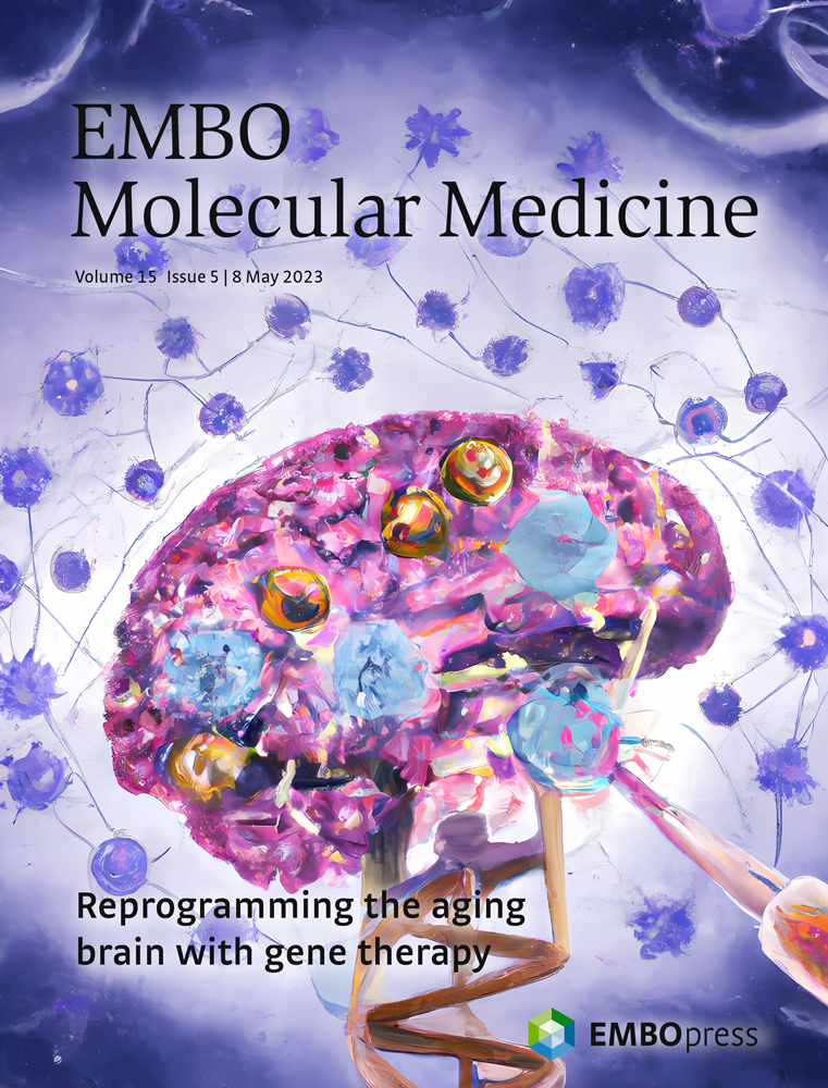
As people age, they often experience problems with their memory and cognitive abilities. This happens in part because their brains become mildly inflamed. But there may be a solution: a small group of special T cells, called regulatory T cells, could help reduce this inflammation in aging brains. Administering a protein called interleukin-2 (IL2) can help these special T cells grow and prevent inflammation. Now, researchers at VIB, KU Leuven, Babraham Institute, and i3S have tested this approach in mice and found that it can prevent neurological decline. Their findings, published in EMBO Molecular Medicine suggest that targeting the immune system might keep people’s brains healthy as they age.

Emanuela Pasciuto, co-first author of the study: “Our goal was to see whether we could slow down the aging process of the brain by changing its immune system through the delivery of IL2. We know that inflammation plays a significant role in various aging processes, and IL2 could help us tilt the balance back in our favor.”

Inflammation in the aging brain

Aging is a degenerative process that affects the whole body, including the brain. As we age, our brains may experience cognitive decline, affecting our memory and ability to think clearly. Increasing evidence suggests that inflammation in the brain, called “inflammaging,” can worsen this decline. Inflammaging is caused by immune cells entering the brain as we age. This inflammation can activate microglia, the resident immune cells in the brain, and induce neuroinflammation, leading to cognitive decline and dementia.

However, researchers have found a way to reduce inflammation in the brain by targeting a small group of special immune cells in the brain called regulatory T cells. Previously, the team of Adrian Liston (VIB-KU Leuven, Babraham Institute) and Matthew Holt (VIB-KU Leuven, i3S Porto) showed that administering a protein called Interleukin-2 (IL2), which helps regulate the immune response, increased the number of regulatory T cells in the brain. This treatment has been successful in mouse models of traumatic brain injury and neuroinflammation.

Now, the researchers want to see if delivering IL2 directly to the brain can help reduce age-induced inflammation and cognitive decline.

Gene therapy improves brain aging

In their latest study, the team discovered that delivering IL2 to the brain improved brain function in aging mice. The research showed that the treatment restored cognitive performance in spatial memory tests, allowing older mice to form new memories almost as well as young mice. The mice given IL2 treatment were better at remembering visual cues than those that did not receive the treatment. Additionally, some of the changes in cellular aging in the brain were reversed, especially among several types of glial cells, which are critical to support overall brain function and health.

Pierre Lemaitre, co-first author of the study: “Our approach was to harness the body’s own system to regulate inflammation and to boost it precisely where it was needed.”

IL2 was delivered to the brain using a gene therapy vector, which is a tool that provides genetic material to specific cells. The additional dose of IL2 allowed the regulatory T cells to survive and create an anti-inflammatory environment. Matthew Holt and Lidia Yshii, co-senior authors of the study: “This reinforces our belief that viral vector-based systems are the way forward for the delivery of therapeutics to combat chronic neurodegenerative diseases and preventing cognitive decline in aging populations.”

Adrian Liston, senior author of the study: “The most important part of this study is the high potential for translation into patients. Inflammation is a process that is conserved in both mice and humans, and regulatory T cells can respond to IL2 in both species. However, there are still regulatory hurdles to clear, and it’s crucial to ensure safety before testing it in patients. Nonetheless, we see a clear path to conducting clinical trials.”

The laboratory is working through spin-off company Aila Biotech to drive entry of this therapeutic into clinical trials. Read the full study here

Saturday
Feb112023

Overnight staining of flow cytometry samples │ Oliver Burton

Monday
Nov142022

Doing more with less: Improving flow cytometry staining

Our latest paper isn't going to cure cancer. But it may just making research into curing cancer 10-fold cheaper. A major cost for immunology, oncology and haematology labs is antibodies, especially with the fancy (and expensive!) new dyes coming on the market, which let us hit the 40-50 parameter range in flow cytometry. Fortunately, Dr Oliver Burton in our lab has developed a simple and easy approach to reduce those costs by 10-fold, while also improving the quality of the data: shifting to overnight staining. You can read the full paper here, describing the protocol, fixatives and optimisation approaches, but essentially, lower antibody concentrations give less background, and if left overnight to stain are able to give even better signal detection. It is also a game-changer for wet-lab staff: rather than having all day dissections and staining, and then cueing for flow cytometer hours in the evening, you can set everything up, put on the staining and get an early night. Next morning your cells are ready to go! 
Out now at Current Protocols!
Thursday
Oct272022

New cause for primary immunodeficiency discovered

Our lab has a new study on primary immunodeficiencies out now at Cellular & Molecular Biology! We studied two families with combined immunodeficiency and found mutations in the Calcium channel ITPR3. The mutations reduce the function of the channel, making the channels 100-fold less capable of initiating a Calcium flux after cellular stimulation. T cells from the patient had poor responses throughout the signalling cascade: reduced Calcium flux, poor nuclear localisation of NFAT1 and reduced proliferative burst, explaining the impeded response to infections. The most severe patient required a bone-marrow transplantation to correct the defect, while the other patient is doing well with regular IgIV treatment. The work established ITPR3 as a new cause of primary immunodeficiency, after previously assuming that these Calcium channels had too much redundancy to be a cause of genetic disease. Read the full paper here, or take a look at the illustrated abstract below for a short-cut summary!
Monday
Jun202022

'Eureka moment' as impact of brain injury in mice reduced

Wednesday
Jun152022

Brain Tregs on The Immunology Podcast

Listen to a great episode of The Immunology Podcast to hear a section on our recent paper about brain Tregs.